Septal Mitral Annular TDI

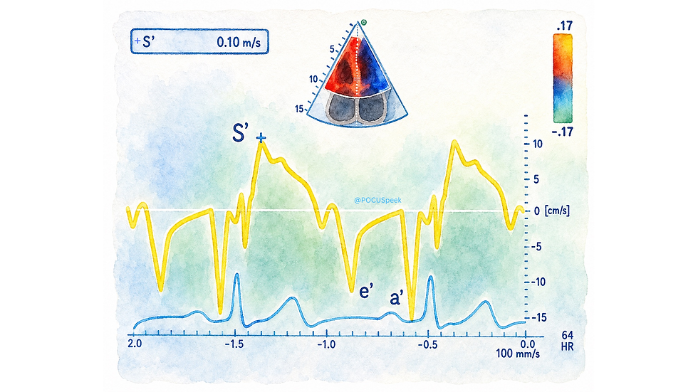
This image is a pulsed-wave tissue Doppler imaging trace of the mitral annulus, from the apical 4-chamber view. It shows the classic annular velocity sequence:
S′ = systolic annular velocity
e′ = early diastolic annular velocity
a′ = late diastolic annular velocity from atrial contraction
S′ — systolic mitral annular velocity
S′ reflects longitudinal systolic shortening of the LV, mainly subendocardial longitudinal fibre function. During systole, the mitral annulus moves toward the apex, generating the S′ wave
In this image, S′ is 0.10 m/s = 10 cm/s. BSE normal reference intervals give lower normal limits for mean mitral annular S′ of approximately ≥6.4 cm/s age 20–40, ≥5.7 cm/s age 40–60, and ≥4.9 cm/s over 60. So an S′ of 10 cm/s suggests preserved longitudinal systolic annular velocity, assuming good alignment and correct sample position
e′ — early diastolic mitral annular velocity
e′ is the key tissue Doppler marker of LV relaxation. It occurs immediately after mitral valve opening, when the LV lengthens and relaxes in early diastole. Physiologically, e′ reflects active myocardial relaxation, recoil and untwisting
The BSE diastolic guideline explains that e′ is inversely related to tau: when relaxation slows, tau increases and e′ falls. Therefore, a reduced e′ is a marker of impaired LV relaxation. Septal e′ is normally lower than lateral e′, so both sites should ideally be measured and interpreted with age-specific reference values
In this image, e′ is the first downward diastolic tissue Doppler wave after S′. The important teaching point is:
Low e′ tells us the LV relaxes slowly. It does not by itself prove raised filling pressure. A patient can have delayed relaxation with low e′ and still have normal LV filling pressure, particularly in older age or early hypertensive heart disease
a′ — late diastolic annular velocity
a′ reflects atrial contraction pulling the mitral annulus in late diastole. It corresponds to the transmitral A wave
In early impaired relaxation, the LV fills less efficiently in early diastole, so atrial contribution increases. Therefore a′ often becomes relatively dominant, and the e′/a′ ratio falls below 1. BSE describes this pattern: with impaired LV relaxation but normal or initially elevated filling pressure, annular e′ falls and e′/a′ often remains <1
But in advanced restrictive physiology, a′ may reduce because the atrium is contracting against a high LV end-diastolic pressure. So a small a′ can mean poor atrial function, AF, high LVEDP, or annular disease depending on context
A simple teaching line: e′ tells us about relaxation; a′ tells us how much the atrium has to rescue late filling
E/e′ — the pressure clue, not the relaxation clue
E/e′ combines transmitral flow with myocardial relaxation
The transmitral E wave is blood-flow velocity from LA to LV in early diastole. The e′ wave is annular relaxation velocity. When relaxation is impaired but filling pressure is normal, both E and e′ tend to fall, so E/e′ may remain normal. But when LV compliance worsens and LA pressure rises, E increases while e′ remains low, so E/e′ rises. BSE states that increased E/e′ is therefore a marker of raised LV filling pressure / LA pressure
BSE cut-offs:
Average septal + lateral E/e′ >14
= highly specific for PCWP >15 mmHg / raised LV filling pressure
E/e′ <8 = specific for normal LV filling pressure
If only one site is valid:
Septal E/e′ >15 suggests raised LVFP
Lateral E/e′ >13 suggests raised LVFP.