RVOT Acceleration Time

Updated: Jul 6
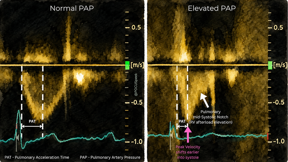
This infographic is showing RVOT pulsed-wave Doppler: flow leaving the right ventricle through the pulmonary valve into the pulmonary artery
The core idea is:
Normal pulmonary vascular bed = low afterload → smooth RV ejection → longer acceleration time
Elevated pulmonary artery pressure / raised pulmonary vascular impedance = high afterload → early peak velocity + shortened acceleration time ± mid-systolic notch
Pulmonary acceleration time / RVOT acceleration time / PAT is the time from the start of RV ejection to the peak systolic velocity in the RVOT Doppler envelope
In normal pulmonary vascular resistance, the RV ejects into a low-resistance, compliant pulmonary arterial tree. Flow accelerates more gradually, reaches peak velocity later, and then decelerates smoothly
In pulmonary hypertension, the RV ejects into a high-impedance circuit. The pulmonary artery pressure rises earlier, reflected waves return earlier, and the RVOT velocity curve peaks earlier. So the acceleration time becomes shorter
BSE does not use RVOT acceleration time as a standalone diagnosis of pulmonary hypertension. It uses it as one of the additional echocardiographic signs that supports the probability of pulmonary hypertension
In the BSE pulmonary hypertension protocol, one of the pulmonary artery category signs is:
RVOT Doppler acceleration time <105 ms and/or mid-systolic notching
On the left infographic, the normal RVOT PW Doppler has a relatively rounded, symmetric-looking systolic envelope, meaning:
low pulmonary artery pressure
good pulmonary artery compliance
low RV afterload
uninterrupted forward flow during systole
The RV can keep ejecting smoothly because it is not fighting an early, steep rise in pulmonary artery impedance
On the right, the peak velocity is reached early. That is the key visual marker.
In elevated pulmonary pressure or raised pulmonary vascular resistance, the RV faces a higher afterload almost immediately after the pulmonary valve opens. The pressure wave travelling into the pulmonary arteries is reflected back earlier. This causes earlier deceleration of forward flow
So instead of a smooth dome, the spectral profile becomes: early sharp acceleration → early peak → rapid deceleration
That shortened acceleration time is why RVOT AT is useful when the TR jet is absent, incomplete, contaminated, or unreliable
The arrowed notch is the most advanced teaching point in the infographic
A mid-systolic notch is a brief reduction or interruption in forward RVOT flow during systole. It is sometimes called a flying W or W-shaped Doppler pattern
Physiologically, it reflects abnormal pulmonary vascular impedance and wave reflection. The RV starts ejecting, but the abnormal pulmonary arterial load pushes back during systole, causing a transient dip in forward velocity. BSE treats mid-systolic notching as equivalent supportive evidence alongside RVOT acceleration time <105 ms
In plain language: the notch is the pulmonary circulation “pushing back” against RV ejection
That is why it is more than a shape. It is a sign of increased RV afterload
BSE starts with TR Vmax where possible
A clearly high TR velocity, especially TR Vmax >3.4 m/s, gives high echocardiographic probability of pulmonary hypertension if the signal is good. If TR Vmax is lower or unavailable, BSE asks you to look for additional signs from different categories. RVOT AT <105 ms or mid-systolic notching sits in the pulmonary artery category of those additional signs
The other categories include right ventricular morphology/function and RA/IVC signs. For example, BSE lists signs such as RV/LV basal diameter ratio >1.0, septal flattening, pulmonary artery diameter >25 mm, IVC dilatation with reduced collapse, and RA area >18 cm²
So the correct BSE logic is:
RVOT acceleration time is helpful, but it is not pure pulmonary pressure. It can be affected by:
tachycardia
low stroke volume
severe RV systolic dysfunction
pulmonary valve stenosis or RVOT obstruction
significant pulmonary regurgitation
poor PW sample position
incorrect measurement to the wrong peak
post-operative congenital anatomy
acute PE versus chronic pulmonary vascular disease
Also, a normal-looking RVOT AT does not fully exclude PH, especially if the rest of the echo or clinical story is worrying
For an acute physician, this trace is especially useful in three situations:
when TR Vmax is absent or poor quality. You still need clues about pulmonary pressure and RV afterload
when the patient may have acute PE or acute RV strain. A short PAT or notching supports increased RV afterload, though it does not diagnose PE by itself
when assessing whether raised TR velocity is believable. If TR Vmax is high and the RVOT Doppler also shows short AT or notching, the physiology becomes much more coherent.