Diastolic Function Approach

Updated: Jul 8
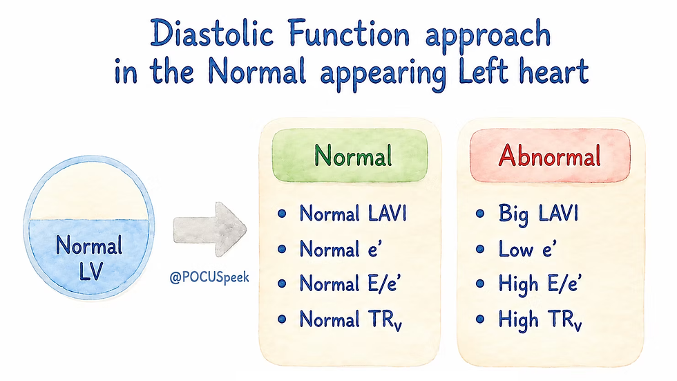
Do not ask: 'normal or abnormal?'
Ask: 'Is relaxation impaired, and are LV filling pressures elevated?'
Core integrated assessment:
1. Myocardial relaxation/recoil
Use septal e′, lateral e′, and age/context. Reduced e′ suggests impaired relaxation, but isolated low e′ is not enough to diagnose raised filling pressure
2. LA remodelling/chronicity
Use indexed LA volume, ideally with LA strain if available. A raised LAVi supports chronic elevation of filling pressure, but can also reflect AF, mitral valve disease, athlete’s heart, ageing, or chronic hypertension
3. LV filling pressure estimate
Use average E/e′, mitral inflow pattern, pulmonary venous flow where helpful, and clinical context. A high E/e′ supports raised LV filling pressure, but it is less reliable in some settings such as significant mitral annular calcification, prosthetic valves, constriction, regional annular dysfunction, and significant MR
4. Pulmonary pressure response
When left atrial pressure / LV filling pressure is chronically or significantly raised, pressure backs up in this direction:
LV filling pressure ↑ → LA pressure ↑ → pulmonary venous pressure ↑ → pulmonary artery pressure ↑ → RV systolic pressure ↑ → TR velocity ↑
So TR velocity is not measuring LV pressure directly. It is telling us that the right side is now “seeing” the consequences of high left-sided pressures through the pulmonary circulation
We can therefore use TR velocity as a downstream marker of elevated left-sided filling pressure, but interpret cautiously. High TRV may reflect pulmonary vascular disease, lung disease, PE, RV pathology, or chronic pulmonary hypertension, not just LV diastolic dysfunction
5. Mitral inflow pattern
Add E/A ratio and E-wave deceleration time. These help grade physiology: impaired relaxation, pseudonormalisation, or restrictive filling. In advanced interpretation, mitral inflow is not optional; it helps explain why the tissue Doppler and LA data look the way they do.
6. Discordance check
If the parameters disagree, do not force a label. Report: 'Diastolic assessment is indeterminate; no convincing evidence of elevated LV filling pressure at rest' or 'Findings suggest impaired relaxation without definite elevation of LV filling pressure.'